OCT is often used to evaluate disorders of the optic nerve as well. OCT has made possible detection of abnormalities of peripapillary retinal nerve fiber layer RNFL in newly diagnosed and chronic IIH.
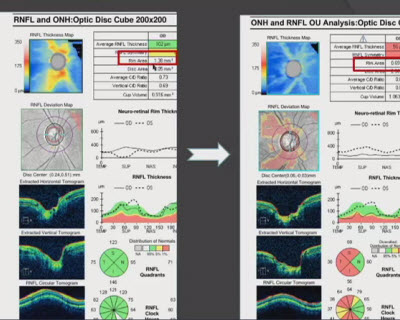
 Optical Coherence Tomography Of The Optic Nerve Head And Retinal Nerve Download Scientific Diagram
Types of Optic Nerve Scanning Technologies OCT Optical Coherence Tomography Devices include.
Oct optic nerve. Optical coherence tomography OCT can aid in the differential diagnosis of optic nerve head ONH elevation. Optical coherence tomography OCT is an imaging technique used widely in the management of macular diseases and glaucoma but it is gaining popularity in neurologic and neuro-ophthalmologic conditions as well. Cirrus HD-OCT RTV ue-100 Spectralis Topcon 3D-OCT 2000 and others CSLO Confocal Scanning Laser Ophthalmoscopy HRT SLP Scanning Laser Polarimetry GDx.
In-Depth Analysis of Nerve Fiber Layer Structures OCT technology is transforming the way physicians assess nerve fiber layer thickness ganglion cell complex thickness and a range of parameters on the optic disc. Specific OCT hallmarks such as V contour and a lumpy-bumpy appearance associated with optic disc edema and optic nerve head drusen respectively were investigated in isolation from line scans added to photographs of various elevated ONHs in a web-based survey distributed among. Since its introduction in Ophthalmology approximately a decade ago the use of this technology has disseminated into the clinical practic.
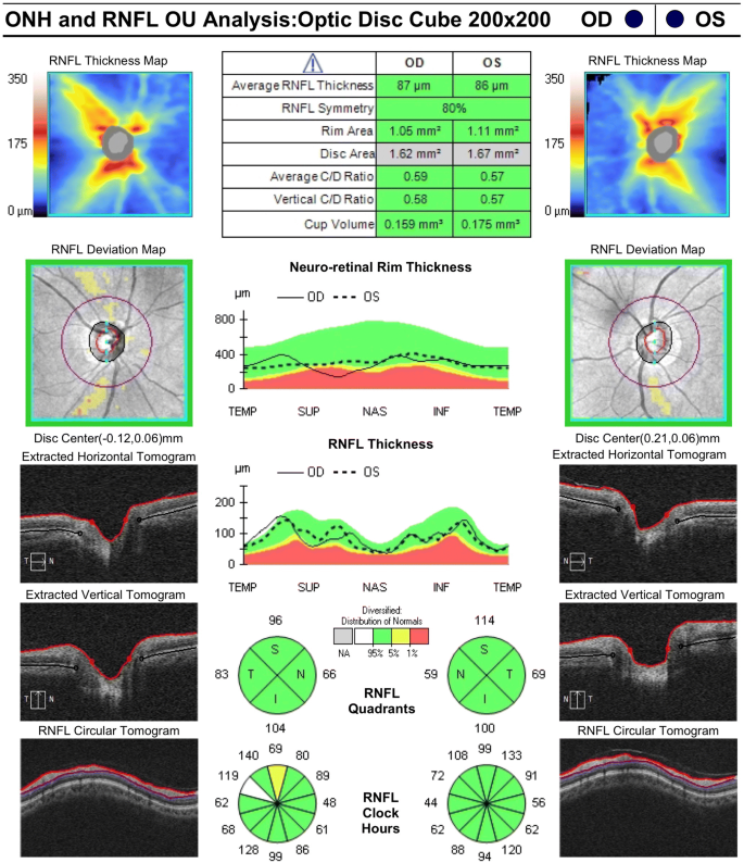
Each of the OCT devices provides the RNFL thickness curve on an age-adjusted normative database where green is considered normal yellow is borderline and red is abnormal RNFL values below the 99th. The optic nerve size changes in early life and is likely stable after age 10. Already indis- pensable for retinal analysis OCT is now becoming essential for glaucoma and other optic neuropathies.
Optical coherence tomography OCT is an easy quick and non-invasive technique using infrared light to obtain in vivo images of macula and optic nerve head ONH close to histological resolution. OCT also provides direct visualisation of the optic nerve. The OCT exam helps your ophthalmologist see changes to the fibers of the optic nerve.
For example it can detect changes caused by glaucoma. Optovue OCT systems utilize advanced software to provide extensive analysis of optic disc structures in simple easy-to-read reports. The major achievement of OCT is by documentation of quantitative changes in the optic nerve head atrophy edema or normal.
Posterior segment evaluation with optical coherence tomography OCT allows visualization of the vitreous retinal layers retinal pigment epithelium RPE and the choroidal layers. OPTICAL COHERENCE TOMOGRAPHY ANGIOGRAPHY OCTA Non-invasive imaging technology that allows in vivo visualization of the retinal and choroidal vasculatures including the peripapillary network. It cannot be used with conditions that interfere with light passing through the eye.
Glaucoma is one of a range of optic neuropathies and OCT also appears useful in the analysis of all diseases of the optic nerve and to some extent the central nervous system when ocular. The optic nerve optic disc optic disk optic nerve head ONH area is approximately 21-28 mm 2 in whites who are not highly myopic depending on the measurement method utilized. This monograph aims to give an update on the use of OCT in glaucoma and pathologies of the optic nerve.
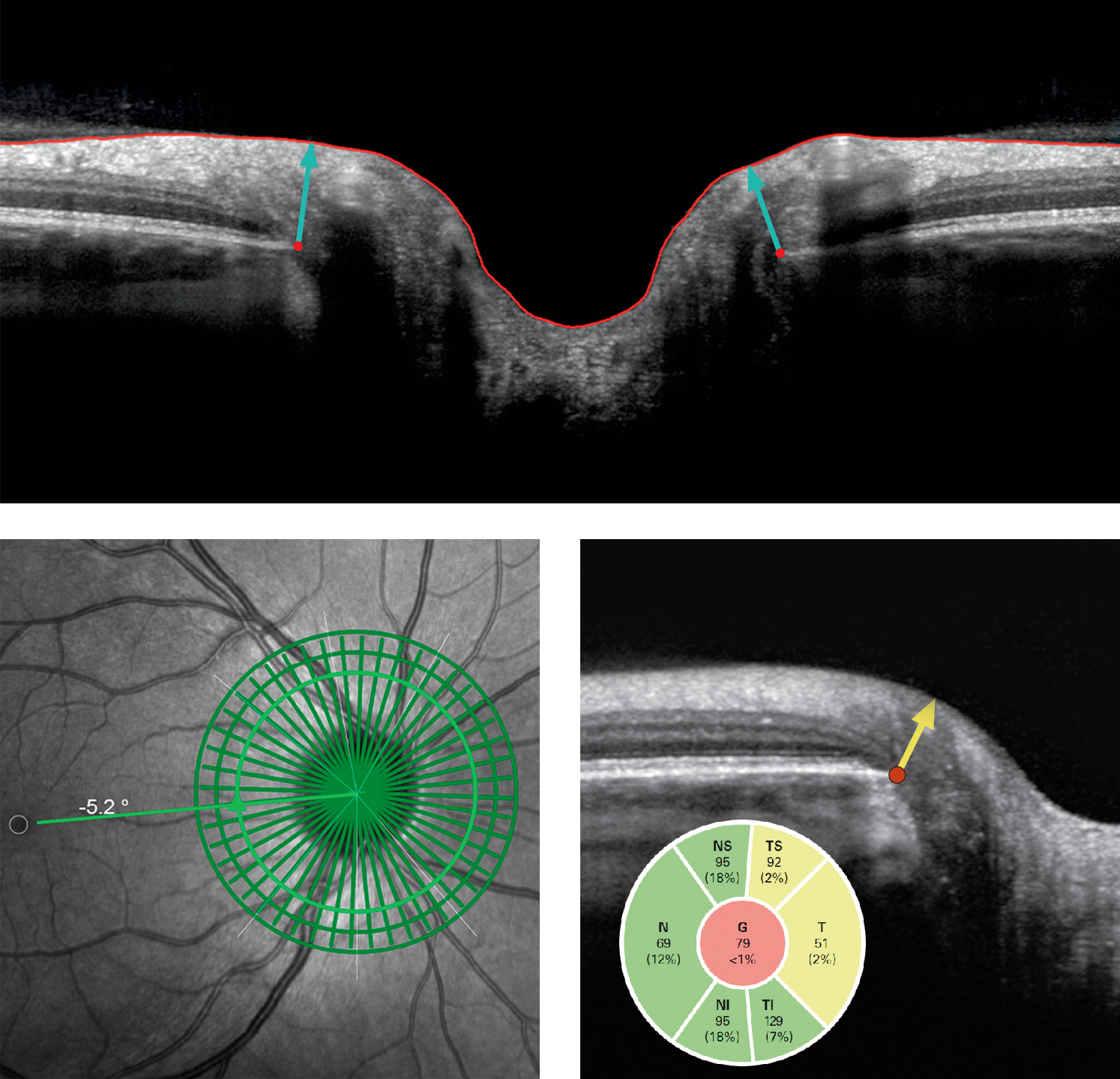
SD-OCT scan of a normal right eye. Optical coherence tomography OCT is a rapid non-contact method that allows in vivo imaging of the retina optic nerve head and retinal nerve fibre layer RNFL. The vitreous retinal layers and choroidal layers are visible.
An OCT scan creates a composite image of the various layers of the optic nerve and retina to allow us to measure their thickness including the thickness of the retinal nerve fibre layer RNFL. Opt 20 066008 2015. The RTVue device scans the optic nerve head with multiple radial and circular scans and generates the RNFL thickness map along a 345-mm diameter circle centered on the optic disc.
OCT relies on light waves.
 Optical Coherence Tomography In Compressive Lesions Of The Anterior Visual Pathway Kaushik Annals Of Eye Science
 Figure 1 From Oct Measurements In Patients With Optic Disc Edema Semantic Scholar
 Diagnosis And Progression Rnfl And Optic Nerve Head Imaging American Academy Of Ophthalmology
 Oct Indispensable In The Diagnosis Of Glaucoma
 Differentiating Mild Papilledema And Buried Optic Nerve Head Drusen Using Spectral Domain Optical Coherence Tomography Ophthalmology
 Diversity In Optical Coherence Tomography Normative Databases Moving Beyond Race Springerlink
Evaluating The Optic Nerve For Glaucomatous Damage With Oct Glaucoma Today
The Impact Of Optic Nerve And Related Characteristics On Disc Area Measurements Derived From Different Imaging Techniques
 The Optical Coherence Tomography Oct Showed Normal Images Of The Download Scientific Diagram
 Conventional Testing Images In A Normal Subject A B Optic Disc Download Scientific Diagram
 The Use Of Oct In Differential Diagnosis Of Elevated Optic Discs The Journal Of Optometric Education
 Heidelberg Engineering Receives Fda Clearance To Market Spectralis Oct Glaucoma Module Premium Edition Business Wire

Comments
Post a Comment